Current projects
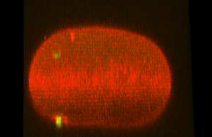
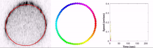
Cytokinetic rings are large actomyosin based structures that promote the physical separation of 2 daughter cells during cytokinesis. Analogous structures are also found in syncytial tissues such as animal germlines where actomyosin based rings termed rachis bridges or ring cannals regulate the cellular openings that connect germ cells and allow for the exchange of cytoplasm. My current work in the lab of Amy Maddox focuses on understanding how cytoskeletal dynamics regulate cytokinetic ring organization and kinetics in dividing embryos as well how cytoskeletal organization controls rachis bridge size, germline architecture and oocyte development.
Quantification of cytokinetic ring organization, remodeling and closure kinetics
|
|
The cytokinetic ring is an incredibly large and complex molecular machine composed of tens of thousand molecules that organize and dynamically rearrange at the cell equator to provide the mechanical force for the physical separation of the two daughter cells during mitosis or meiosis. The molecular regulation of contractile ring assembly is fairly well understood, but the nature of the cytoskeletal rearrangements and dynamics that promote cytokinetic ring contraction largely remain to be elucidated. More specifically, It is currently unknown how individual cytokinetic ring component are organized within the closing ring, how this organization changes as the ring changes size and how this translates into force generation to separate the two daughter cells.

|
I have developed novel imaging approaches that allow for the visualization cytokinetic ring dynamics in dividing cells with unprecedented spatial and temporal resolution. We combine this 4 dimensional high spatial‐ and temporal resolution imaging with custom quantitative image analysis pipelines to study these dynamics. This work allows as to follow local and global changes in composition and kinetics
|
of the closing cytokinetic rings in one C.elegans embryos as well as cells of the embryo at different developmental stages with different size and cell fates.
The role of GCK-1/CCM-3 in regulating cortical contractility during cytokinesis
|
|
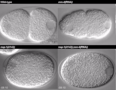
The small GTPase RhoA is a central regulator, activating cortical actomyosin contractility during cytokinesis and other cellular events. It acts as a molecular switch that can alternate between an active and inactive state. The sterile20‐family serine/threonine kinase GCK‐1 and its cofactor CCM‐3 form a conserved complex and suppress RhoA
|
activity in vertebrates. We and others found GCK‐1/CCM‐3 enriched on the stable actomyosin rings of germline intercellular bridges and cytokinetic rings. We showed that GCK-1/CCM-3 participate in a negative feedback loop among RhoA and its cytoskeletal effectors to inhibit contractility. GCK-1 and CCM-3 are recruited by active RhoA and anillin to the cytokinetic ring, where they in turn limit RhoA activity and contractility. This is evidenced by increased RhoA activity, anillin and nonmuscle myosin II in the cytokinetic ring, and faster cytokinetic furrowing, following depletion of GCK-1 or CCM-3. GCK-1 or CCM-3 depletion also reduced RGA-3 levels in pulses and increased baseline RhoA activity and pulsed contractility during zygote polarization. Together, our results suggest that GCK-1 and CCM-3 regulate cortical actomyosin contractility via negative feedback.
Bell KR*, Werner ME*, Doshi A, Cortes DB, Sattler A, Vuong-Brender T, Labouesse M, Maddox AS. Novel cytokinetic ring components drive negative feedback in cortical contractility, Mol Biol Cell. 2020 Jul 15;31(15):1623-1636. (*co-first author) Pubmed
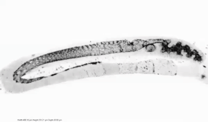
The role of the syncitial germline in oocyte formation
Animal embryogenesis depends on dis-proportionally large oocytes that contain the biomass necessary for cleavage divisions and morphogenesis before the animal accesses extracellular nutrients. Many germlines promote the formation of large oocytes by allowing the exchange of cytoplasm among cell-like compartments of a syncytium. The oogenic syncytial germline of C. elegans is an elongated C-shaped tube in which peripheral germ cell compartments surround a central core of common cytoplasm, the rachis. Germ cells open to the rachis via intercellular bridges (rachis bridges) in their apical surface. Cytoplasm flows from nurse-cell like germ cells near one end of the germline towards and into a subset of germ cells that enlarge and eventually become oocytes. Both the rachis bridges and the apical domain of germ cells that make up the lining are enriched for components of the cortical actomyosin cytoskeleton. It has been suggested that oogenesis is regulated by both rachis bridge size as well as overall contractility of the rachis lining. How these two processes coordinate to promote oogenesis, is not fully understood.
|

|

|
I study how the distribution of conserved structural and regulatory elements of the actomyosin cytoskeleton, on rachis bridges and germ cell comparment membranes affect germ cell structure and oocyte formation. To test whether these protein abundance dynamics represented significant increases and decreases, and how abundance dynamics correlate to germ cell development, we used SiZer (SIgnificant ZERo crossing of the derivatives) statistical analysis to identify germline regions with statistically significant changes in rachis bridge perimeter. Subdividing the germline into distinct regions based on the changes in rachis bridge size allows us to identify significant correlations between change in protein distribution and rachis bridge size. Our observations suggest that regulation of germline architecture and oogenesis rely on a balance of distribution of contractile
|
components on rachis bridges and rachis lining, as well as other factors including cytoplasmic flows.
Rehain-Bell K, Love A, Werner ME, MacLeod I, Yates JR 3rd, Maddox AS. A Sterile 20 Family Kinase and Its Co-factor CCM-3 Regulate Contractile Ring Proteins on Germline Intercellular Bridges. Current Biol. 2017 Mar 20:26(7): p860–867 Pubmed
Rehain-Bell K, Love A, Werner ME, MacLeod I, Yates JR 3rd, Maddox AS. A Sterile 20 Family Kinase and Its Co-factor CCM-3 Regulate Contractile Ring Proteins on Germline Intercellular Bridges. Current Biol. 2017 Mar 20:26(7): p860–867 Pubmed
Past Research
Cortical actomyosin dynamics during cytokinesis

|
During my PhD research in the laboratory of Michael Glotzer I used high resolution confocal microscopy to study cytoskeletal dynamics during establishment of embryonic polarity and cytokines in C.elegans embryos. My research involved developing an experimental approach to genetically separate two redundant pathways involved in cytokines in the same embryo. This approach revealed that these two pathways promote drastically different organization of the actomyosin cytoskeleton both sufficient to drive furrow ingression. Furthermore this research contributed to the identification and characterization of the roles of three different proteins in regulating cortical actomyosin dynamics, spindle positioning and cytokinesis in the one cell C.elegans embryo.
|
1. Werner M, Munro E, and Glotzer M. Astral Signals Spatially Bias Cortical Myosin Recruitment to Break Symmetry and Promote Cytokinesis. Current Biology 2007 Aug 7;17(15):1286-97 Pubmed
2. Tse YC*, Werner ME*, Longhini KM, Labbé JC, Goldstein B, Glotzer M. RhoA activation during polarization and cytokinesis of the early C. elegans embryo are differentially dependent on NOP-1 and CYK-4. Mol Biol Cell. 2012 Oct;23(20):4020-31 (*co-first author) Pubmed
3. Afshar K, Werner ME, Tse YC, Glotzer M, Gönczy P. Regulation of cortical contractility and spindle positioning by the protein phosphatase 6 PPH-6 in one-cell stage C. elegans embryos. Development 2010 Jan;137(2):237-4 Pubmed
Ciliogenesis and Cilia orientation
Ciliated epithelia are found throughout nature, performing locomotor functions in a variety of aquatic organisms and providing directed fluid flow in a variety of developmental and physiological contexts. The loss of directed fluid flow in humans can result in hydrocephaly, situs inversus, infertility, and respiratory dysfunction. The epithelium of Xenopus laevis embryos is covered with multiciliated cells, which generate a robust flow oriented from anterior to posterior. This system has proven useful for dissecting the role that planar cell polarity (PCP) signaling and hydrodynamic forces play in orienting motile cilia.

|

|

|
Role for the Cytoskeleton in Cilia orientation


|
During my work as a Postdoc in the lab of Brian Mitchel we showed that intracellular effectors interpret polarity to organize cellular morphology in accordance with asymmetric cellular function. We observe that both cellular actin and microtubule networks undergo drastic reorganization, providing differential roles during the polarized organization of cilia. Using computational angular correlation analysis of cilia orientation, we report a graded cellular organization downstream of cell polarity cues. Actin dynamics are required for proper cilia spacing, global coordination of cilia
|
polarity, and coordination of metachronic cilia beating, whereas cytoplasmic microtubule dynamics are required for local coordination of polarity between neighboring cilia. Additionally, I contributed to the characterization of novel proteins required for cilia orientation.
1 Werner ME, Hwang P, Huisman F, Taborek P, Yu CC, Mitchell BJ. Actin and microtubules drive differential aspects of planar cell polarity in multiciliated cells. J Cell Biol. 2011 Oct 3;195(1):19-26. Pubmed
2 Chien YH, Werner ME, Stubbs J, Joens MS, Li J, Chien S, Fitzpatrick JA, Mitchell BJ, Kintner C. Bbof1 is required to maintain cilia orientation. Development 2013 Aug;140(16):3468-77 Pubmed
The microtubule associated Protein CLAMP in MCC intercalation and cilia polarity

|
The directed movement of cells is a fundamental aspect of tissue morphogenesis. The skin of Xenopus laevis embryos represents an excellent model to study the movement of cell across epithelia. Multiciliated cells (MCCs) and ionocytes (ICs) are specialized cell types that differentiate in a subapical layer of the epidermis. These cells then move in a directed manner toward the outer epithelial cells where they undergo radial intercalation pushing through cell vertices and establishing new junctions while maintaining epithelial integrity. We identified a novel function for both the microtubule-binding protein CLAMP and members of the microtubule-regulating Par complex during intercalation.
|
Specifically, we show that Par3 and aPKC promote the apical positioning of centrioles, whereas CLAMP stabilizes microtubules along the axis of migration. We propose a model in which the Par complex defines the orientation of apical migration during intercalation and in which subcellular localization of CLAMP promotes the establishment of an axis of microtubule stability required for the active migration of cells into the outer epithelium.
Werner ME, Mitchell JW, Putzbach W, Bacon E, Kim SK, Mitchell BJ. Radial intercalation is regulated by the Par complex and the microtubule-stabilizing protein CLAMP/Spef1. J Cell Biol. 2014 Aug 4;206(3):367-76 (Journal cover article) Pubmed
Werner ME, Mitchell JW, Putzbach W, Bacon E, Kim SK, Mitchell BJ. Radial intercalation is regulated by the Par complex and the microtubule-stabilizing protein CLAMP/Spef1. J Cell Biol. 2014 Aug 4;206(3):367-76 (Journal cover article) Pubmed
Other cilia related projects

|
Multiciliated cells represent an interesting variation of centriole duplication in that these cells generate greater than 100 centrioles, which form the basal bodies of their motile cilia. This centriole amplification is proposed to require a structure termed the deuterosome, thought to be capable of promoting de novo centriole biogenesis. I contributed to the characterization of the deuterosome and identify it as a site for the
|
localization of Cep152, Plk4, and SAS6. This study also identified CCDC78 as a centriole and deuterosome associated protein that is essential for centriole amplification.
Klos Dehring DA , Vladar EK*, Werner ME *, Mitchell JW, Hwang P, Mitchell BJ Deuterosome mediated centriole biogenesis. Developmental Cell 2013 Oct 14;27(1):103-12. (*co-second author) Pubmed
Klos Dehring DA , Vladar EK*, Werner ME *, Mitchell JW, Hwang P, Mitchell BJ Deuterosome mediated centriole biogenesis. Developmental Cell 2013 Oct 14;27(1):103-12. (*co-second author) Pubmed

|
Primary ciliary dyskinesia (PCD) is a genetic disorder in which impaired ciliary function leads to chronic airway disease. Exome sequencing of a PCD subject identified a frameshift variant that introduces a stop codon in amino acid 308 of the growth arrest-specific protein 2-like 2 (GAS2L2). In this research, using human nasal cells, mouse models, and X.laevis embryos, we show that GAS2L2 is abundant at the apical surface of ciliated cells, where it localizes with basal bodies, basal feet, rootlets, and actin filaments. Loss of GAS2L2 in mouse tracheal epithelial cell (mTEC) cultures and in X. laevis
|
embryos leads to defects in cilia cilia orientation, cytoskeletal orientation and defects in cilia mediated fluid flow.
Bustamante-Marin XM, Yin W-N, Sears PR, Werner ME, Brotslaw EJ, Mitchell BJ, Jania CM, Zeman KL, Rogers TD, Herring LE, Refabert L, Thomas L, Amselem S, Escudier E, Legendre M.8, Grubb BR, Knowles MR, Zariwala MA, and Ostrowski LE. Lack of GAS2L2 causes a new variant of PCD by impairing cilia orientation and mucociliary clearance. Am J Hum Genet 2019 Feb 7;104(2):229-245 Pubmed
Bustamante-Marin XM, Yin W-N, Sears PR, Werner ME, Brotslaw EJ, Mitchell BJ, Jania CM, Zeman KL, Rogers TD, Herring LE, Refabert L, Thomas L, Amselem S, Escudier E, Legendre M.8, Grubb BR, Knowles MR, Zariwala MA, and Ostrowski LE. Lack of GAS2L2 causes a new variant of PCD by impairing cilia orientation and mucociliary clearance. Am J Hum Genet 2019 Feb 7;104(2):229-245 Pubmed

|
C21orf59/Kurly (Kur), a cytoplasmic protein with some enrichment at the base of cilia, is needed for motility, proper cilia polarization in the zebrafish kidney and the larval skin of Xenopus laevis. CRISPR/Cas9 coupled with homologous recombination to disrupt the endogenous kur locus in Xenopus resulted in the asymmetric localization of the PCP protein Prickle2 being lost in mutant multiciliated cells. Kur also makes interactions with other PCP components, including Disheveled. This supports a model wherein Kur plays a dual role in cilia motility and polarization.
|
Jaffe KM, Grimes DT, Schottenfeld-Roames J, Werner ME, Ku TS, Kim SK, Pelliccia JL, Morante NF, Mitchell BJ, Burdine RD. c21orf59/kurly Controls Both Cilia Motility and Polarization. Cell Rep. 2016 Mar 1;14(8):1841-9. Pubmed
Regulation of microtubule dynamics

|
AMBMP is a small molecule that has been previously reported to be both a Wnt agonist and a microtubule (MT) regulator. We showed that a detailed analysis of AMBMPs effects on MTs and on MT associated cellular processes including cell polarity, ciliogenesis, and cell migration. Specifically, treatment of Xenopus embryos with AMBMP leads to defects similar to the MT depolymerizing drug nocodazole, including a failure to generate or polarize
|
cilia (depending on the timing of treatment) and a loss of the cell movements associated with radial intercalation. The dramatic effect AMBMP has on basic MT based cellular functions suggests that its usefulness as a Wnt regulator is questionable. Moreover, it may be an important new tool for experimental or pharmacological manipulation of MTs.
Werner ME, Del Castillo U, Ventrella R, Brotslaw EJ, Mitchell BJ. The small molecule AMBMP disrupts microtubule growth, ciliogenesis, cell polarity and cell migration. Cytoskeleton (Hoboken). 2018 Oct;75(10):450-457. Pubmed
Werner ME, Del Castillo U, Ventrella R, Brotslaw EJ, Mitchell BJ. The small molecule AMBMP disrupts microtubule growth, ciliogenesis, cell polarity and cell migration. Cytoskeleton (Hoboken). 2018 Oct;75(10):450-457. Pubmed

|
MT-associated proteins (MAPs) have context-dependent roles in regulating their stability and dynamics. The poorly characterized clamp/Spef1 gene encodes a MAP and is expressed in several tissues and cell types. We have shown that CLAMP dynamically interacts with the microtubule latice and promotes promotes centriole positioning and radial intercalation of multiciliated cells by connecting microtubule organization to apico-basal polarity.
|
1. Werner ME, Mitchell JW, Putzbach W, Bacon E, Kim SK, Mitchell BJ.. Radial intercalation is regulated by the Par complex and the microtubule-stabilizing protein CLAMP/Spef1.. J Cell Biol. 2014 Aug 4;206(3):367-76 Pubmed
2. Kim SK*, Zhang S*, Werner ME*, Brotslaw EJ, Mitchell JW, Altabbaa MM, Mitchell BJ. CLAMP/Spef1 regulates planar cell polarity signaling and asymmetric microtubule accumulation in the Xenopus ciliated epithelia. J Cell Biol. 2018 May 7;217(5):1633-1641 (*co-first author) Pubmed
2. Kim SK*, Zhang S*, Werner ME*, Brotslaw EJ, Mitchell JW, Altabbaa MM, Mitchell BJ. CLAMP/Spef1 regulates planar cell polarity signaling and asymmetric microtubule accumulation in the Xenopus ciliated epithelia. J Cell Biol. 2018 May 7;217(5):1633-1641 (*co-first author) Pubmed

|
Mamalian cylicin is localized to the cytoskeletal calyx that surrounds part of the nucleus in the head of sperm. The two highly conserved proteins, CYLC-1 and CYLC-2 in C.elegans have been suggested to function in the intracellular compartmentalization of tubulin levels in uterine muscle cells. We generated CRISPR tagged worm strains for both genes and show that both proteins are highly enriched in sperm in both hermaphrodites and males concentrated in puncta presumed to be membranous organelles.
|
Krauchunas AR , Werner ME, Britt N, Chen DS, Maddox AS, Singson A. C. elegans CYLC-2 localizes to sperm. MicroPubl Biol 2020 Sep 29;2020:10.17912/micropub.biology.000314. Pubmed
